Preface: Sulphuric ether entered the surgical armamentarium on October 16, 1846, the day Dr. John Collins Warren risked his reputation on an experimental substance to blunt the pain of his scalpel while excising the facial tumor of his patient, Edward Gilbert Abbott (1825-1855). The providential Abbott operation has inspired many artists, writers, and photo historians, but their interpretations of Ether Dome Daguerreotype No. 1 – the Hawes daguerreotype claimed to represent the operation – have accreted around a good deal of guesswork. It is generally conceded that Abbott is not the patient in the photograph, but the prevailing view that he is an actor posing as Abbott for a staged "Reenactment daguerreotype" commemorative is also doubtful. There is scant evidence that verifies the identities of the doctors or their motivations in posing for EDD No. 1, and the more the reenactment construct is studied, the more inconsistencies in the details that emerge. Photographic records of the first ether operations were eventually made, this is known, and Hawes is established as the photographer. Dr. Warren's place in history was secured when, according to the legend, he turned to the spectators and declared, "Gentlemen, this is no humbug," but was this one of the many seeds of fiction planted that day? And is it possible the first photograph of ether anesthesia represents an actual surgery in which Dr. Warren is neither playing the lead role, nor even among the subjects who are posing for the camera?
I propose that the first photograph of etherization captures not a reenactment but rather an enactment, one of the actual ether surgeries prosecuted at Massachusetts General Hospital within the first months following the Abbott operation. Drawing from my experience as a visual artist, this will be a physiognomic study of the confusion that surrounds the identities and mischaracterizations of Ether Dome Daguerreotype No. 1 as a staged reenactment, a kind of fictionalized tableau vivant. The identities of the principals will be examined, including that of the etherized patient, and correctives offered. I also draw upon the valuable guidance I have received through my correspondence with scholars who are intimate with the Ether Dome daguerreotypes and, as this is a work in progress, guidance I hope to acquire in the future.
Completed edits for all pages. Added content to the section on Charles Bertody, including a jpeg of his portrait painted by Bernard Zakheim for the Toland Hall murals, UCSF Parnassus Campus.
Uploaded revisions to the section on Henry Jacob Bigelow.
Finally cataloged Ether Dome Daguerreotypes Nos. 3-5 in the bibliography, representing Dr. John Collins Warren's exploratory surgery of a diseased fibula in a young Portuguese sailor. Again, these daguerreotypes are incredibly important images in the bibliography and warranted a considerable amount of research to get the cataloging right, after historians have gotten the attributions so wrong.
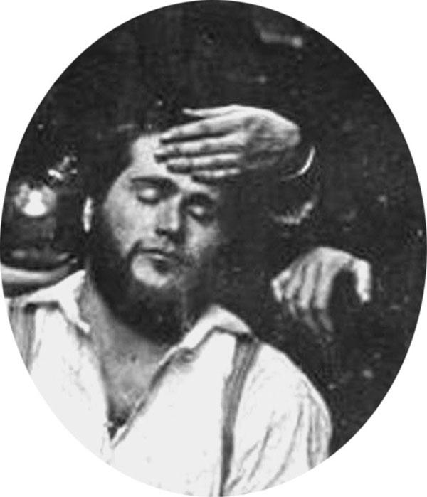
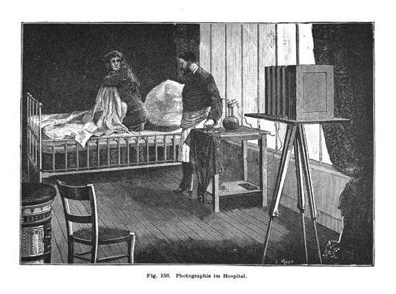
It is not at all unusual for nineteenth century medical photographs to come with little or no documentation of their subject matter. Even so, given the vast resources of the Getty it is a little surprising that they would get the description of an object in their archives wrong, especially for one as important as "Ether Dome Daguerreotype No. 4," taken by Boston photographers Albert Sands Southworth & Josiah Johnson Hawes (link: »»). This historic image records one of the early surgeries conducted with ether anesthesia, but it is misidentified: John Collins Warren is the operator, not Dr. Townsend. Furthermore, the photograph was taken in July, not "late spring" as claimed by the Getty and so it couldn't possibly be a record of "the retiring Dr. Warren's last lecture," since Harvard Medical College was not in session.
The patient was a 19-yr-old Portuguese sailor named Francis Manuel, who went under Warren's knife on July 3, 1847 for exploratory and intervention surgery of diseased fibula in his left leg (link: »»). It was the first of the three photographs of an ether surgery taken that day, not the second, and the date of the photo session can now be established with certainty. The archival records of Massachusetts General Hospital should have the exact time of day, and possibly additional details not found in Warren's extensive notes on the operation.
After many months of research, I am finally able to add four of the five Ether Dome Daguerreotypes to my bibliography of nineteenth century medical photography, beginning with the attribution of Parkman's treatment of dislocated humerus, depicted by Ether Dome Daguerreotype No. 1, the misidentified "Reenactment Daguerreotype" of Southworth and Hawes. The daguerreotypes are the most significant images in the bibliography, and it was necessary to get their cataloging right, after historians have gotten them so wrong.
Completed Appendix 1 for the ether anesthesia paper. Information on the "Boston Society for Medical Improvement" photograph.
Updated Oettinger on exophthalmos.
Added a few lines of text to the summation, regarding Dalton and his role in the ether anesthesia scenario.
Completed the rewrites of the John Call Dalton section.
Fixed the caption to the comparative views of William T. G. Morton. Added trace evidence of Morton's height that refutes the claim he is the etherizer in the reenactment photograph. There is a photograph of Morton with his family in which he appears quite tall, as opposed to the shorter height of the etherizer, who is most likely John Call Dalton.
Reworked parts of the Introduction.
Added information on a another possible attribution for Figure 1 as Francis Parkman (1823-1893). This Parkman was an eminent historian of the American wilderness, notably publishing work on the Oregon Trail. He was not a physician, but an argument for the attribution can be made based on the appearance of Daniel Denison Slade in the photograph. The two men were very close friends at the time and remained so for their entire lives. Slade accompanied Francis during his first explorations of the forests of New England.
The patient was a 19-yr-old Portuguese sailor named Francis Manuel, who went under Warren's knife on July 3, 1847 for exploratory and intervention surgery of diseased fibula in his left leg (link: »»). It was the first of the three photographs of an ether surgery taken that day, not the second, and the date of the photo session can now be established with certainty. The archival records of Massachusetts General Hospital should have the exact time of day, and possibly additional details not found in Warren's extensive notes on the operation.
Rewrote part of the description for Jonathan Mason Warren. No new content.
Reworked the first and last paragraphs of the Summation section. Mostly grammar, no new content.
Reworked the segment covering Parkman in the Southworth and Hawes daguerreotype.
Added text and corrected grammar in the EDD No. 2 segment, thought by some historians as representing the treatment by actual cautery of the patient Athalana Golderman. But it is more likely that the daguerreotype represents the etherization of a patient undergoing surgery for epulis on July 3, 1847.
Reworked the Henry Jacob Bigelow segment a little and corrected an error in the date of the referenced table.
Uploaded revisions on the Southworth & Hawes Ether Dome Daguerreotypes Nos. 3-5.
Uploaded revisions on Ether Dome Daguerreotype No. 2.
Uploaded revisions to the Summation section.
Revisions to John Call Dalton, the etherizer in Ether Dome Daguerreotype No. 1 He collaborated with Bigelow after the Abbott operation to suss out the secret of Morton's obtunding nostrum (Letheon). Prepared the tables of all the ether anesthesia surgeries at Massachusetts General Hospital published in 1848.
Completed the revisions to the section on the Robert Cutler Hinckley painting of the Abbott operation.
Finished the section on the April 3 scenario of the reenactment daguerreotype.
Partial completion of Jonathan Mason Warren's second report, pending additional information.
Completed and uploaded Jonathan Mason Warren's first report.
Additional revisions to the identities in the Ether Dome "reenactment" daguerreotype. Uploaded descriptions for Drs. Samuel Parkman, George Parkman, Daniel Denison Slade, and John Call Dalton. The link above brings up the title page and index in a new tab.
Completed the revisions to the Henry Jacob Bigelow section in the Ether Dome daguerreotype paper.
Completed the revisions to the Jonathan Mason Warren section in the Ether Dome daguerreotype paper.
Uploaded the revised "Reenactment" section to the Ether Dome daguerreotype paper.
Uploaded the revised "Introduction" to the Ether Dome daguerreotype paper.
Began uploading the documentary material for the first photographs of ether surgery. This includes identities of the surgical team represented by Ether Dome Daguerreotype No. 1. The link only brings up the Preface and Index page. More pages will be added shortly.
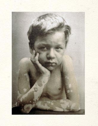
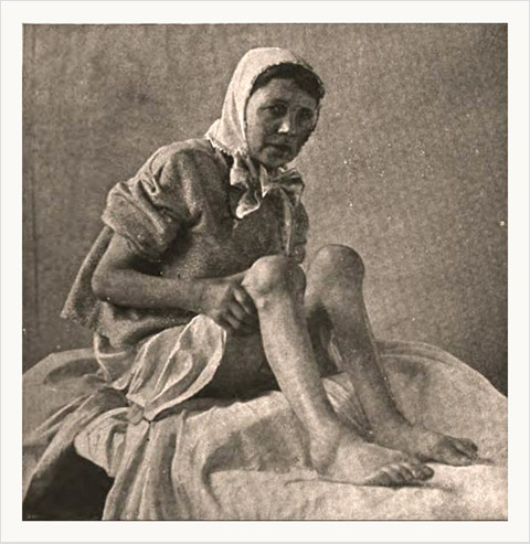
This photograph of leprosy affecting a young male subject comes from John Wood's book of poems titled,
Endurance & Suffering. The book was reviewed by the Canadian
Medical Association Journal—the print version of the journal has 65,000 subscribers and the on-line version gets
over a million hits a month. To access the PDF file of the review, click on "Begin manual download" on the web page
here »»
An expanded version with more images and poems can be accessed directly
here »»
Cataloged the Carl Ernst Baer elephant folio on Blumenbach taxonomy. Titled, "Types principaux des différentes races humaines dans les cinq parties du monde. Modelés sous la diréction du Pr. Baer de St. Petersbourg."
Cataloged Dr. George Hinckley Lyman's eulogy of Charles Edward Buckingham.
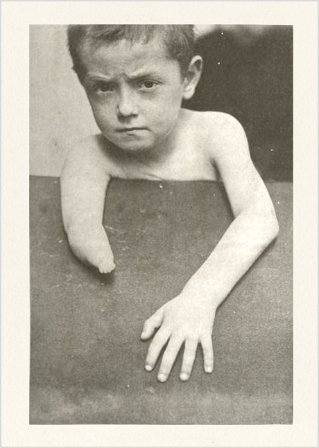
Cataloged Dr. Arthur Breese Stout's surgical report on a child titled, "Successful operation on harelip." The photo is an early example of an in-text use of photographic illustration.
Cataloged John Henry Dunn's case report on leprosy titled, "A clinical lecture on leprosy."
Cataloged John Newport Langley's contribution to the Goltz vs. Ferrier dispute over cerebral localization, titled, "Report on the parts destroyed on the right side of the brain of the dog operated on by Prof. Goltz."
Cataloged a monograph by the Prussian military physician, Bernhard von Beck. Titled, "Ueber die Wirkung moderner Gewehrprojektile insbesondere der Lorenz'schen verschmolzenen Panzer-Geschosse auf den thierischen Körper." The photographic material is unverified until I find a copy.
Updated a doctoral thesis on basal encephalocele written by Dr. Max Sperling and titled, "Ein Fall von beiderseitigem Hirnbruch an den innern Augenwinkeln bei einem Neugeborenen, nebst Bemerkungen über die an dieser Stelle vorkommenden angeborenen Bildungsfehler."
Cataloged a report by Dr. James Hardie on Tagliacotian flap procedure for treating burn cicatrix and contraction.
Cataloged a report by Dr. Kelburne King on excision of the scapula in an 8 year-old subject.
Cataloged a case report by Dr. John Forrest Dewar on placenta previa.
Cataloged a rare copy of Dr. Abner Otis Kellogg's seminal writings on a Shakespearean system of psychology, titled, "Shakspeare's delineations of insanity, imbecility, and suicide."
Cataloged a rare hospital report for San Giacomo in Rome title, "Resoconto morale amministrativo dello ospedale di S. Giacomo in Augusta nel triennio 1871-1873." Written by Dr. Alessandro Angelucci.
The significance of this day, 9/11, is not lost. Cataloged the Königlich Preussischen Kriegsministeriums 10 in 8 volume set of medical and surgical reports from the Franco-Prussian war of 1870/71. "Sanitäts-Bericht über die Deutschen Heere im Kriege gegen Frankreich 1870/71)."
Cataloged two volumes of orthopedic appliances published by Gustav Ernst. "Orthopćdic apparatus. A series of illustrated plates, with corresponding descriptions of the various forms of mechanism employed in the treatment and cure of the numerous deformities of the human body."
Cataloged Dr. Leonard William Sedgwick's report on the Cheselden and Mead busts commissioned for the new building construction of St. Thomas's Hospital, "The Old Students' Gift. 1871."
Cataloged a paper on gunshot wounds to the lower extremities by William MacCormac, "Some remarks on gunshot wounds of the lower extremity."
Cataloged a surgical case of Dr. Roderick Maclaren of Carlisle, England, titled, "On a case of periosteal excision of the head and part of the shaft of the humerus."
Revised and enlarged Friedrich Bezold's paper on vascular corrosion casts, "Die Corrosions–Anatomie des Ohres. Festschrift der Julius-Maximilians-universität zu Würzburg zur feier ihres 300 jährigen bestehens gewidmet von dem Ärztlichen verein in München. "
Cataloged Friedrich Bezold's paper on facial paralysis, "Labryinthnecrose und Paralyse des Nervus facialis."
Cataloged Friedrich Bezold's second treatise on mastoiditis titled, "Erkrankungen des Warzentheiles."
Cataloged Friedrich Bezold's first treatise on mastoiditis titled, "Die Perforation des Warzenfortsatzes vom anatomischen Standpunkte aus."
Updated "Atlas des menschlichen Gehörorganes," by Dr. Nicolaus Rüdinger.
Cataloged Hugo Wilhelm von Ziemssen's speech celebrating the opening of the Klinishe Institute in Münich. Titled, "Ueber die Aufgaben des klinischen Unterrichts und der klinischen Institute : Rede gehalten bei der Eröffnung des Med.-Klinischen Institutes der Königl. Universtität München am 8. Juni 1878 : Nebst einer Beschreibung d. klinischen Institutes zu München." It is a limited run of the edition with the added photo, probably distributed to the benefactors of the research center.
Cataloged Dottore Fabio Grilli's monograph on the application of Paquelin's thermocautery in surgical cases. Titled, "Sistema antisettico e termo-cauterio. Osservazioni pratiche."
Cataloged a paper by the eminent Harvard University anatomist, Thomas Dwight, titled, "The significance of bone structure."
Updated and corrected the catalog entry for Profeta's "Sulla Lepra" and added a better jpeg.
Cataloged a monograph on cerebral circulation, written by two Naples University physiologists, Gaetano Rummo and his nephew, Andrea Ferrannini. Title of the work is, "La circulation cérébrale chez l'homme ŕ l'état normal et sous l'influence des substances hypnogčnes."
Cataloged a doctoral thesis submitted by Paul Lavis, an obscure Lyon physician, titled, "De la cheiloplastie par le procédé du pont sus-hyoďdien, avec ilot mentonnier d'arręt, pour la restauration des grandes pertes de substance de la lčvre inférieure."
Updated the catalog entry for James Aitken Meigs memoriam written by Hamilton. Added the albumen portrait.
Cataloged a four page tract titled, "An appeal in behalf of a hospital for the University of Pennsylvania." No date, no publisher, and no author name recorded, however it is well established that William Pepper–future provost of the University of Pennsylvania–was the author. It is quite satisfying to find a photographic work by this scholar to add to this bibliography.
Cataloged, "Cannes et son clima," by Jules-Edmond-Théophile de Valcourt.
Cataloged Ernst von Bumm's treatise on the gonococcus bacterium, "Der Mikro-Organismus der gonorrhoischen Schleimhaut-Erkrankungen, Gonococcus–Neisser."
Cataloged another paper by Van Duyse titled, "Éléphantiasis de la paupičre supérieure."
Cataloged van Duyse's paper, "Cryptophtalmos."
Cataloged "Ueber die gemeinschaftlichen Ursachen von Glaucom, Myopie, Astigmatismus und den meisten Cataracten," by Wilhelm Roeder.
Cataloged an early statistical study on climate and endemic disease. Very rare. "Estudios médicos sobre el clima de la Provincia de Jujui," by Dr. Ismael Carrillo.
Cataloged Hermann Welcker's monograph, "Schiller's Schädel und Todtenmaske, nebst Mittheilungen über Schädel und Todtenmaske Kant's."
Cataloged Ranney's paper, "The treatment of functional nervous diseases by the relief of eye-strain."
Cataloged a paper on treatment of rotary lateral curvature of the spine written by Dr. Reginald Sayre, the son of the orthopedic surgeon Lewis Sayre.
Cataloged Dr. Robert William Taylor's paper, "Lichen ruber as observed in America and its distinction from lichen planus."
Cataloged a sequel to Dr. French's first paper, "On a perfected method of photographing the larynx."
Cataloged a landmark paper, "On photographing the larynx," by Dr. Thomas Rushmore French.
Cataloged another paper by Dr. William Thompson Lusk on ectopic pregnancy.
Cataloged "A case of rupture of the uterus and a case of Cæsarean section," by Dr. William Thompson Lusk.
Cataloged a paper on an exanthema caused by rhubarb by Dr. Herman Goldberg.
Cataloged a paper on leprosy in the Sandwich Islands and Mexico by Dr. Prince Albert Morrow.
Another paper by Dr. Milton Josiah Roberts, published in the New York Journal of Medicine and titled (in part), "Spina bifida, chronic internal hydrocephalus, hydrorrhachis, double convergent strabismus, and double talipes varus, existing concurrently."
Cataloged a paper on the wire corset for the treatment of spinal deformities, published by Dr. Milton Josiah Roberts.
Cataloged an encomium on Louis Pasteur, published with a photograph of a bust of Pasteur. "Discurso pronunciado por el alumno Don Ricardo Mandado y respuesta dada por el Dr. Rodríguez Méndez."
Cataloged a paper on the collotype process, submitted by a physician photographer: "De quelques applications de la photographie ŕ la médecine," by Pierre Bernard (1859–1889). A promising career that was cut too short.
Cataloged a paper on photographing bone structures with polarized light. "Untersuchungen über das Verhalten des Knochengewebes im polarisirten Lichte," by Dr. Viktor von Ebner.
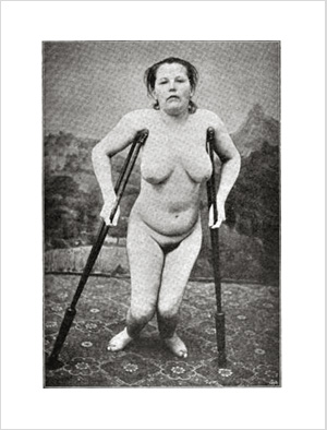
Very few photographs of myositis ossificans published in the nineteenth century: "Demonstration eines Falles von Myositis ossificans progressiva," written by Oswald Kohts of Strassburg.
Catalogued a paper on the utility of the intrauterine sponge tampon for treating placenta previa. Written by Hermann Jungbluth of Aachen and titled, "Zur Behandlung der Placenta prćvia."
Finally got around to cataloging Joseph Gibert's study on rickets. Titled, "Quels rapports peuvent exister entre le rachitisme et la syphilis."
Yet another memorial. This one honoring Raffaele Piria, the doctor who discovered salicylic acid. Written by his protégé, Stanislao Cannizzaro: "Sulla vita e sulle opere di Raffaele Piria, discorso."
Cataloged still another memorial, this one for Dr. Jacob Herz, who broke through barriers of institutional racism at Erlangen School of Medicine to become its first Jewish professor.
Cataloged Raphaël-Horace Dubois's study on the pyrophorus beetle. His discovery of the remarkable luciferin-luciferase system continues to find new applications in biochemistry research, especially for tagging reporter genes in genetic studies. Title: "Les Élatérides lumineux: contribution a l'étude de la production de la lumičre par les ętres vivants."
Cataloged another memorial, this one for the beloved health officer of Rochester, NY., Dr. William Francis Sheehan.
Cataloged a memorial for a beloved Portland, Maine, physician. "John Taylor Gilman, M. D., Portland Maine: A Memorial for the Family," by Charles Henry Bell.
Cataloged a collection of reports from the surgical clinic of Antonino D'Antona in Naples. Dr. D'Antona contributed a chapter, but most of the book was written by his protégé, Giovanni Pascale. Title of the book, "Osservazioni di patologia e clinica chirurgica sui casi piů importanti della clinica."
Cataloged a paper by Friedrich Louis Hesse, founder of the first German school of dentistry (Leipzig). Titled, "Ueber die Muskeln der menschlichen Zunge," his doctorate for surgery.
Cataloged a paper by Walter Brewster Platt titled, "Hypertrophy of clitoris; amputation."
Cataloged a paper by John Fenton Evans titled, "Cases of muscular atrophy and degeneration." Evans died from the malaria bacillus he was researching.
Cataloged a second paper by Edward Long Fox titled, "Enlargement of the Spleen."
Cataloged a paper by Edward Long Fox titled, "Case of spontaneous cure of spina bifida, followed by hydrocephalus."
Updated the description for an important document on leprosy titled, "Leprosy and Segregation ; with Photographs," by Archdeacon Henry Press Wright. Several of the images are the work of Dr. Henry Vandyke Carter (1831-1897), the artist/physician who created the images for Gray's Anatomy.
Cataloged a memorial of a bright young New England surgeon, who died in the early days of the Civil War. "Ebenezer Kimball Sanborn; a memorial," by Dr. Samuel Burnham.
Cataloged a paper on the pathogenesis of glaucoma, titled, "A further investigation of the pathology of glaucoma," by Dr. Priestley Smith.
Cataloged a paper on the surgical treatment of strabismus, titled, "Advancement of Rectus," by Dr. Arthur Edward Prince.
Updated Dr. George Henry Savage on myxoedema.
Paper by Dr. Julian John Chisolm on Blepharoplasty.
Cataloged Dr. George Harley's sensational report on a postmortem delivery by caesarean section.
Cataloged Izett Anderson's paper on neurofibromatosis titled, "On Molluscum Simplex."
Updated the catalog description for Buckminster Brown's paper on a case of double congenital malformation of the hip.
Cataloged a memoir of Dr. John Maclean, first professor of chemistry at Princeton.
Cataloged a doctoral thesis on fat embolism syndrome titled, "Beiträge zur Fettembolie," submitted by Alfred Halm.
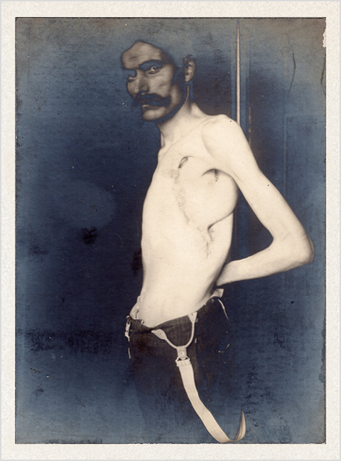
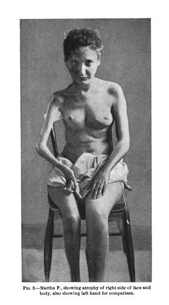
There were only about 60 cases of Hemiatrophia facialis on record when this doctoral thesis was written by Hermann Graff. Titled, "Ein Fall von Hemiatrophia facialis progressiva verbunden mit neuroparalytischer Ophthalmie."
Cataloged a report on a very rare disease, namely, Progressive hemifacial atrophy (PHA). The report was written by two young residents at St. Bartholomew's Hospital in London. Title: Two cases of hemiatrophia facialis. Authors: Walter Hamilton Hylton Jessop and Oswald Auchinleck Browne.
Cataloged an album of clastic models from the collection of Paris urolologist, Jean-Baptiste-François Mallez. Title of the album is, "Album d'anatomie pathologique." The wax models were made by Jules Talrich (1826-1904) and photographed by Léon Crémičre, (1831-1913).
Updated Barwell on club foot.
Cataloged a monograph on suspension as a treatment for scoliosis titled, "The homśopathic treatment of spinal curvatures according to the new principle." Written by Edward Carroll Franklin.
Cataloged a paper in Swedish on the morphological changes caused by childbirth on the vulva and vagina. Titled, "De endnu efter involutionsperiodens slutning märkbare tegn pĺ en forudgĺet födsel og betydningen af samme ved retsmedicinske undersögelser," written by Emil Rode.
Cataloged a very rare offprint of a paper titled, "An unique case of multiple exostoses," written by the nephew of Joseph Leidy.
Cataloged a paper titled, "On the treatment of cleft palates in infants, and on the cure of clefts of the hard and soft palates by operation under chloroform," written by Sir Thomas Smith.
Cataloged a paper titled, "Observations on the angle of the neck of the thigh-bone," written by Sir George Murray Humphry.
Cataloged another doctoral dissertation, this one on excision of the elbow joint for suppurating tumor. By Henry Lucas-Fontaine and titled, "Étude sur la résection appliquée ŕ la tumeur blanche du coude." His son was also a physician and became a member of the French resistance.
Updated Vaucher description.
Cataloged what is possibly the first published photograph of Dr. Max Schede's operation for empyema. Illustrating the doctoral dissertation of Johannes Thomée and titled, "Iets over empyeembehandeling." Below is another photograph of Schede's procedure, taken in 1905:
Cataloged an early work on the treatment of obesity. Written by Jean-François Dancel, titled, "Hygične; nouveaux préceptes pour diminuer l'embonpoint sans altérer la santé."
Cataloged an early work published in Japan. Written by Suart Eldridge, titled, "Notes on the crania of the Botans of Formosa."
Cataloged Jean-Lčo Testut, "Recherches anthropologiques sur le squelette quaternaire de Chancelade (Dordogne)."
Cataloged Frédéric Gross on a paper, titled, "Contribution ŕ l'histoire des corps libres articulaires dans l'arthrite déformante"
Cataloged the offprint of a paper titled, Observaçőes de um caso de hermaphrodismo masculino, with three photographs taken by the author, Dr. Carlos May Figueira of the Lisbon Medical School. Figueira modernized the curriculum of the school with courses in microscopy, photomicrography, and ophthalmoscopy.
Cataloged the Italian translation of Bernard Perez's, La psychologie de l'enfant. L'enfant de trois ŕ sept ans, with an introduction and notes written by Lombroso.
Cataloged Chevers's textbook on forensic medicine, A manual of medical jurisprudence for India..etc.
Finally completed the cataloging of Lombroso's work, L'uomo delinquente.
Cataloged a paper by Serafino Varaglia & Bernardino Silva, Note anatomiche ed antropologiche sopra 60 crani e 42 encefali di donne criminali italiane.
Cataloged Lombroso's paper, I pazzi criminali.
Catalogued Lombroso's paper on the epileptiform physiognomy.
Third Lombroso work with photographs. Titled, "Fisionomia delle donne criminali," also co-written with Dr. Antonio Marro.
Second Lombroso work with photographs. The "German album," co-written with Dr. Antonio Marro.
Finally tackling Lombroso. On the occasion of the Outsider Art Show here in NYC, here is possibly the first published photograph relevant to the art of the insane. Illustrating a paper by Lombroso and co-authored with the French photographer, Maxime du Camp. Titled, L'arte nei pazzi.
Catalogued a paper on cyclopia, from the examination of a specimen, written by Émile-Marie Valude and Gustave Vassaux.
Catalogued a paper on a sarcoma of the ocular adnexa, written by the Swiss-born ophthalmologist, Edmund Landolt, and his protégé, Samuel Eperon. Title, "Sarcome de la région interne de la paupičre gauche; extirpation; autoplastic."
Catalogued a paper on tumor of the lacrimal gland, reported by Victor de Britto of Porto Alegre, Brazil.
Catalogued a paper by Jakob Stilling on myopia and its association with the morphology of the orbit.
Catalogued Samuel-Jean Pozzi's biography of Paul Broca with its portrait by Pierre Petit.
Catalogued Escherich on his discovery of E. coli and its action.
Photograph by Charles Workman, M.D., for a paper on hemilateral atrophy in a case of scleroderma, written by John Lindsay Steven of Glasgow, and published in the 1897 issue of International Clinics: A Quarterly of Clinical Lectures (vol. ii., »»). Workman prepared the microphotographs that illustrated Escherich's seminal paper on intestinal bacteria.
Catalogued a paper written by the Russian dermatologist, Alexis Pospeloff.
Catalogued another photographic atlas of skin diseases. By Van Haren Noman, the first part was published in 1889, just squeaking into the bibliography.
Catalogued a paper on ophthalmic instruments written by Anatole-Pierre-Louis Gillet de Grandmont, titled, "Périoptométrie et chromatopsie; périmčtre et chromatoptomčtre."
Catalogued a commemorative of Dr. Francesco Secondo Beggiato, written by Paolo Lioy.
Catalogued a doctoral thesis on scoliosis, written by Pierre Jaune Henri de Bruďne and titled, "Over het onderzoek en de behandeling van scoliosis," illustrated with photographs by the author.
Catalogued a doctoral thesis written by an obscure physician titled, "De la cachexie pachydermique (myxoedčme des auteurs anglais)."
Catalogued a report published in, "Medical reports upon the character and progress of leprosy in the East Indies, being answers to interrogatories drawn up by the Royal College of Physicians, London." The report is a thorough response to a list of queries about leprosy in the Pooree district of India, and was submitted by James John Durant. It is the only report in the book that is illustrated with photographs. Stereographs, specifically.
Catalogued a paper titled, "Remarks on a case of traumatic facio-brachial monoplegia," written by Mackintosh Alexander Thomas Collie, a surgeon in the Indian Medical Service (IMS).
Catalogued a paper titled, "A case of monstrosity; multiple pregnancy," written by Rustomjee Naserwanjee Khory.
Catalogued a third paper by the ophthalmic surgeon Charles Bell Taylor titled, "Abstract of a clinical lecture on certain modifications of the operation for internal and external squint, with a simple method of treating aggravated cases of the latter condition."
Catalogued a hospital report for the Pennsylvania Hospital, published in 1880 by Drs. Thomas George Morton and William Hunt. The plate is a wonderful study of three amputees, photographed by Frederick Gutekunst.
Catalogued a work on malarial fever published by Simon-Antoine-Édouard Burdel and titled, "Simon-Antoine-Édouard Burdel"
Catalogued Benedict Stilling's three volume work, plus atlases, on the morphology of the brain, titled, "Untersuchungen über den Bau des kleinen Gehirns des Menschen."
Catalogued a pamphlet by Tadeusz Zulinski on the comparative morphology of the human brain titled, "Quelques mots au sujet du cerveau d'un Grand-Russe : envoye a la Société d'Anthropologie de Paris par la section anthropologie de la Société des sciences naturelles de Moscou."
Catalogued Jules-René-Martial Brossard's doctoral thesis titled, "Étude clinique sur une forme héréditaire d'atrophie musculaire progressive débutant par les membres inférieurs."
Catalogued Baron Shibasaburo Kitasato's paper on the tetanusbacillus.
Catalogued a paper by Wilhelm His titled, "Ueber Präparate zum Situs viscerum mit besonderen Bémerkungen über die Form und Lage der Leber, des Pankreas, der Nieren und Nebennieren, sowie der weiblichen Beckenorgane," an absolute treasure in 19th century medical literature.
Catalogued a paper on diaphragmatic hernia titled, "Nuove osservazioni intorno all' ernia diaframmatica esposte alla r. Accademia medica di Roma," by Filippo Scalzi.
Catalogued a paper on stenosis of the trachea caused by goiter, written by Hermann Askan Demme, and titled, "Fortgesetzte Beobachtungen über die compressiven Kropfstenosen der Trachea." Possibly the first appearance of photographs in a German medical journal.
Catalogued a landmark paper on the function of neural structures in the skin, written by Alfred Goldscheider and titled, "Neue Thatsachen über die Hautsinnesnerven."
Catalogued a paper on the treatment of facial scarring from skin disease, by Oscar Lassar, "Beitrag zur Narbenverbesserung, mit Krankenvorstellung."
Catalogued a dissertation on the membranous labyrinth by Christian Utz titled, "Beitrag zur Histologie der häutigen Bogengänge des menschlichen Labyrinthes."
Catalogued a dissertation on the signs of epilepsy on the retina by Georges Pichon, titled, "De l'épilepsie dans ses rapports avec les fonctions visuelles."
Catalogued a folio of three large format photographs commissioned by Hubertus Cornelius Antonius Leopoldus Fock to support his writings on aesthetic symmetry.
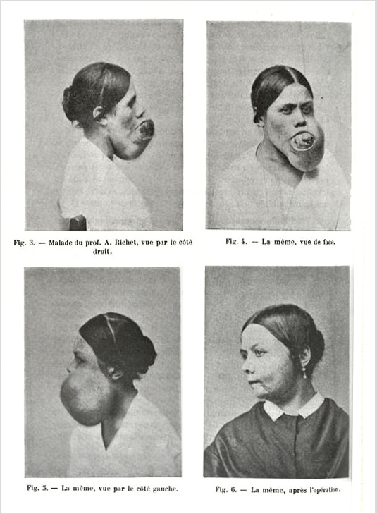
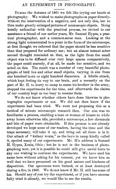
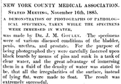
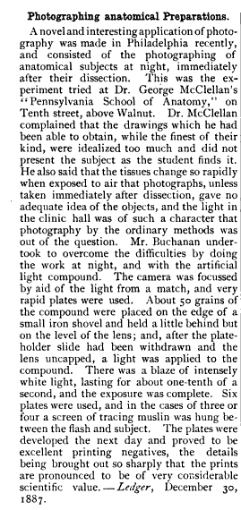
Ces photographies (fig. 3, 4 et 5) portent la signature de M. Pierre Petit, le photographe parisien bien connu. Je suis donc allé aux informations auprčs de lui, et ses deux fils sont tombés d'accord pour me dire que la malade avait été photographiée par eux-męmes, en 1867, dans le service du professeur A. Richet, ŕ la Pitié. Ils ont aisément retrouvé les clichés d'aprčs lesquels ces photographies avaient été faites et j'ai pu ainsi en obtenir des épreuves nouvelles, qui ont servi ŕ faire les gravures ci-jointes. Bien plus, MM. Pierre Petit ont retrouvé un quatričme cliché, dont il n'existe aucune trace au Musée Dupuytren et qui représente la malade aprčs l'opération (fig. 6).—Blanchard, Raphaël Anatole Émile (1899), "Archives de parasitologie," vol. 2, p. 335 »»
Catalogued a paper written by another obscure Italian dermatologist, Amedeo Marianelli. Title of the paper is, "Contributo allo studio del pemfigo vegetante."
Catalogued a paper on Alibert's disease written by a somewhat obscure Italian dermatologist, Lorenzo Mannino. Title of the paper is, "Sulla micosi fungoide di Alibert."
Updated the description for John Curwen, "The original thirteen members of the Association of Medical Superintendents, of American Institutions for the Insane."
Catalogued a treatise on excision of the knee-joint by Peter Charles Price.
Reading on William Harvey is always enlightening, in this case, a Harveian Oratory delivered by Arthur Farre, an analysis on "Harvey's exercises on generation."
Catalogued the eulogies for Joseph Gensoul titled, "Notice sur le docteur Joseph Gensoul."
Catalogued a doctoral thesis by Charles-Alexandre-Gaston Petiau titled, "Contribution ŕ l'étude du traitement du bec de ličvre double compliqué."
Catalogued Hansen on partial albinism, "Einige Beiträge zur Casuistik des Albinismus partialis."
Catalogued Mansurov, "Klinicheskii sbornik po dermatologii i sifilologii. "
Catalogued a paper in the American Journal of Insanity, titled, "Some of the uncommon causes of imbecility," by the British asylum superintendent, Fletcher Beach
Thomas Barnes Hitchcock, "Report on dental histology and microscopy."
James Leon Williams—"On certain disputed points in the development and histology of the teeth."
Catalogued a small atlas on the microbiology of adipose tissue compiled by William Robert Weisiger, founder and president of the Richmond Microscopical Society and titled, "A paper on the relations of the minute blood-vessels to the fat cells in the fascia of the calf's neck."
Catalogued Henry Winchester Sawtelle's operation titled, "Caries of the os calcis; excision; mortification."
Catalogued Edoardo Porro's operation titled, "Dell' amputazione utero-ovarica come complemento di taglio cesareo."
Catalogued a case of vitiglio in a 59 year-old Italian male actor. Title is "Caso di cloasma cacheticum con vitiligine in un vecchio." Discovered no biographical material for the author, Amedeo Thurman.
Catalogued Augustus Choate Hamlin's case of "Molluscum fibrosum."
Catalogued Thomas Taylor Minor's case of rupture of the omentum and double diaphragmatic hernia.
Began writing the description for "Photographs illustrating rare books in the National Medical Library," with plates by Joseph Janvier Woodward and preface notes by John Shaw Billings.
Catalogued Schmedicke's monthly periodical "Der Zahnarzt."
Catalogued a piece of ephemera celebrating the fiftieth anniversary of the Bailličre publishing house titled, "La cinquantaine d'un libraire."
Catalogued a notice necrologique for Jean-Charles-Philippe-Joseph Delvaux de Fenffe.
Catalogued another work by naval surgeon, Jules Roux, titled, "Leçon de cliniquč chirurgicale sur la désarticulation du coude."
Updated Jules Roux's contribution on the internal plantar flap technique for amputation of the foot, titled, "Sur l'amputation tibio-tarsienne."
Cataloged Julius von Michel's study on the decussation of the optic nerve, titled, "Ueber Sehnerven-Degeneration und Sehnerven-Kreuzung."
Cataloged a piece of photographic ephemera titled, "Das Denkmal für Julius Cohnheim auf dem neuen Johannisfriedhofe zu Leipzig. Zur Erinnerung," celebrating the unveiling of the Julius Cohnheim monument in the Leipzig cemetery. Lead author was Wilhelm His.
Cataloged Georges-Marie Félizet's doctoral thesis titled, "Recherches anatomiques et expérimentales sur les fractures du crâne," with its phototypes by Charles Bilordeaux, son of pioneer photographer, Adolphe Bilordeaux.
Updated the description for William Francis Fluhrer's paper titled, "An operation for the extraction of a pistol-ball from the brain through a counter-opening in the skull." Added the following amusing anecdote to the description:
The photographs were taken by George Gardner Rockwood, who introduced the carte-de-visite format to America—the medium of his great success and fame in life. It is fun to speculate how his photographs of Dr. Fluhrer's patient might have inspired a medical hoax, "Brain pictures," that Rockwood wrote and published in the New York Tribune the following year (1887). His story made the claim that he participated in the autopsy of a mysterious german born philologist, Count Borenski. Using a microtome he purportedly acquired from the distinguished microscopist, Dr. John William Schmidt Arnold (1846–1888), Rockwood shaved off tissue samples from a bulge in the Broca region of the fictitious Borenski brain. From these shavings he prepared a sliver bromide photomicrograph that proved too grainy, but next he prepared an albumen plate of sufficient clarity to reveal a vermiform mass of legible characters from Ethiopic, ancient Syriac, and Phoenician languages.
Cataloged a one-page paper by Oliver Fairfield Wadsworth, titled, "Photograph of the fovea centralis of the retina."
Cataloged another paper by Giovanni Calderini, titled, "Esportazione dell' utero dalla vagina; narrazione di un caso seguito da decesso e studii sulla operazione," also illustrated by two dyed albumen prints.
Updated the Jackman and Webster paper on photographing the fundus oculi.
Cataloged a paper by Giovanni Calderini, titled, "Una cretini ed una microcefala nell' Istituto ostetrico di Parma; nota clinico-anatomica," illustrated by two rough, but wonderful dyed albumen prints.
Cataloged Paul von Bruns on induced myxoedema, title, "Zur Frage der Entkropfungs-Cachexia."
Cataloged Léon Defontaine's procedure for restoring mobility to the elbow-joint, titled,"Ostéotomie trochléiforme; nouvelle méthode pour la cure des ankyloses osseuses du coude."
Cataloged another paper on comparative craniology, this time of indigenous siberians, titled, "Ostiacchi e Samoiedi dell' Ob.," by Stefano Sommier
Cataloged a work on comparative craniology by Paolo Mantegazza and Ettore Regalia, titled, "Studio sopra una serie di erani dei Fuegini."
Cataloged a work on comparative craniology by Paolo Mantegazza and Ettore Regalia, titled, "Nuovi studi craniologici sulla Nuova Guinea."
Cataloged Albert Hoffa, "Ueber die Folgen der Kropfoperationen.".
Cataloged Gustave Imbert's doctoral thesis on restoration of the lower lip: Étude sur la restauration de la lčvre inférieure, suivie de la description d'un nouveau procédé pour refaire le bord libre au moyen d'un lambeau.
A Genčve également, nous voyons, chez M. Julliard, une jeune fille qui a été opérée huit jours auparavant pour un goitre (thyroďdectomie du lobe droit); incision verticale linéaire, la seule que fasse jamais Julliard; la réunion est déjŕ parfaite; le drain a été retiré lors du dernier pansement il y a trois jours. Dans ce cas, il n'y avait pas d'indications spéciales. Julliard nous dit qu'il a fait 62 thyroďdectomies, il nous montre l'album renfermant la photographie de ses malades avant et aprčs l'opération. Il nous fait ŕ ce propos la démonstration de sa pince destinée ŕ faciliter les ligatures dans les extirpations de goitre. Le principe consiste ŕ soulever entre les mors écartés de la pince le vaisseau ŕ lier : une ligature est placée en dehors de chacun de ces mors; l'artčre est ensuite secé tionnée dans l'intervalle des mors, c'est-ŕ-dire entre deux ligatures; on est ainsi assuré de laisser sur chacun des segments du vaisseau un bout artériel d'une longueur suffisante pour que la ligature ne glisse pas.—Pages 945-946, Revue de chirurgie, Volume 7.
Updated Edmund Lesser's textbook on skin disease.
Catalogued a possible unicum with two albumen portraits of the infamous Dr. Edward William Pritchard, the "Poisoning Philanderer."
Catalogued Ignazio Zani's necrology of the italian alienist, Antonio Galloni, superintendent to San Lazzaro.
Catalogued Hart on rupture of the peritoneal portion of the vagina during labor.
Catalogued The pathology and treatment of diseases of the skin, written by the colorful character, John Laws Milton, founder of St. John's Hospital for diseases of the skin, and inventor of painful penal collars for treating nocturnal emission.
Catalogued Cesare Belluzzi's paper on induced labor titled, Centuria parti primaturi artificiali provocati dal Dott. Cesare Belluzzi dal 1860 al 1882.
Catalogued Adolf Lorenz's monograph on the treatment of scoliosis titled, Pathologie und Therapie der seitlichen Rückgrat-Verkrümmungen.
Catalogued Johann Bernhard Aloys von Gudden's monograph on the development of cranial sutures, Experimental-Untersuchungen über das Schædelwachsthum.
Catalogued Alexander Macalister's paper, Further evidence as to the existence of horned men in Africa.
Catalogued Henri-Edmond Robiquet's textbook on photography titled, Manuel théorique et pratique de photographie sur collodion et sur albumine.
Catalogued Thomas Scott Lambert's monograph on drowning titled, They are not dead!
Catalogued Robert Battey's case of double monster.
Catalogued Vincenzo De Giaxŕ on using culture plates as photographic negatives: Ueber eine einfache Methode zur Reproduction.
Catalogued one more paper by Nocard, titled Note sur la mammite gangréneuse des brebis laitičres (vulgo: araignée, mal de pis). This completes the cataloguing of the Annales Pasteur.
Catalogued Verujski's paper on the culture of Tricophytan, Recherches sur la morphologie et la biologie du trichophyton tonsurans et de l'achorion Schśnleinii.
Catalogued Roux's paper, La photographie appliquée ŕ l'étude des microbes.
Sur une mammite contagieuse des vaches laitičres. Title of a paper by Nocard and Mollereau on the isolation of Streptococcus agalactiae, originally associated only with bovine mastitis, it has since become the leading neonatal pathogen.
Roux on the flasks he designed for cultivating anaerobic microbes, titled, Sur la culture des microbes anaérobies.
Continued cataloguing the Annales de l'Institut Pasteur photographic material with this paper by Nocard and Roux on cultivation of the tubercle bacillus.
Edmond-Isidore-Étienne Nocard's landmark discovery of the Nocardia asteroides bacterium. The paper is titled, Note sur la maladie des bśufs de la Guadeloupe connue sous le nom de farcin, published in 1888 and catalogued with two plates of heliotypes.
Catalogued another paper on composite photography of skulls, written by the Scottish anatomist, Arthur Thomson.
Updated John Shaw Billings and Washington Matthews papers on composite photography of skulls.
Catalogued a paper on the microcephalus, Manghi Abramo, reported by Ettore Baistrocchi.
Cranio e cervello di un idrocefalo di 19 anni, is the title of a paper by Augusto Tamburini with two views of the skull of a hydrocephalic idiot. The postmortem description is lengthy and incredibly detailed.
Considered by scholars to be a landmark in medical photography, the first published photographs of the fundus oculi were submitted by Dr. William Thomas Jackman and J. D. Webster to the [London] Photographic News in 1885 and 1886. Title of their paper: On photographing the retina of the living human eye.
Catalogued a monograph by Henri Viallanes titled, "La photographie appliquée aux études d'anatomie microscopique," illustrated by a superb photolithograph of the brain of a larval fly. The photo was produced in the workshop of André-Nicolas Quinsac in Toulouse, France.
Catalogued a monograph on embryogenesis of the spine (chick) by Karl Wilhelm von Kupffer, illustrated by the photograms of Berthold-Adolph Benecke.
Catalogued a rare journal paper by Rodolfo del Castillo y Quartiellerz on encephaloid tumor of the face.
Updated Stokes biography on Robert James Graves.