Journal : Royal London Ophthalmic Hospital Reports ; vol. x.
London : J.& A. Churchill, 1880-82.
Description : 25–43 p., [7 l.] pl. ; ill.: 14 photo. figs., 1 tbl. ; 22.5 cm.
Photographs : mounted woodburytypes, reproducing drawings of specimens.
Subject : Eye — Glaucoma ; pathogenesis.
Notes :
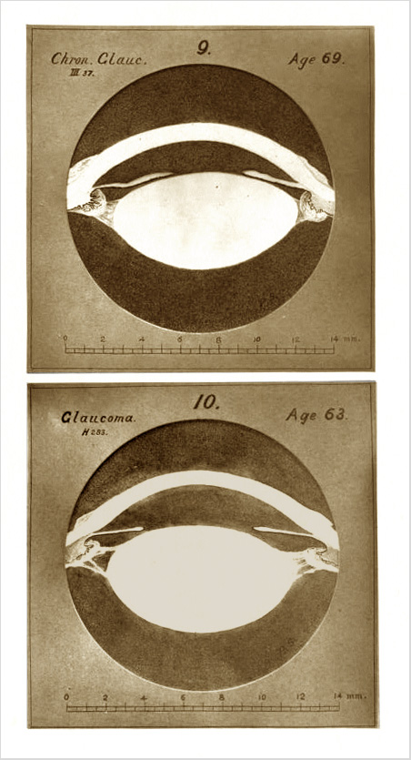
The following was the mode of examination: — The specimen, after lying for seven days unopened in Muller's fluid, was divided by a circnlar incision, corresponding roughly with the ora serrata. The anterior portion, containing the lens and ciliary processes intact, was placed in solution of hydrate of chloral 1 in 20. The remnants of the vitreous humour and hyaloid membrane were carefully removed. The diameter of the lens was measured in several meridians by means of finely pointed spring compasses and a magnifying glass, as the specimen lay in the fluid. Some days later, during which time the lens underwent no perceptible change of size, the specimen was placed in a small covered china dish, containing just enough thick gum-water to cover it, and at once frozen solid by immersion of the vessel in a mixture of ice and salt, and then divided into halves by a meridional section, and returned to the chloral solution. The thickness of the lens was then measured with compasses. Placed with the cut surface looking upwards, the specimen was readily examined with a low power of the microscope as it lay in the chloral solution. By means of a special arrangement of the microscopic camera, and a vertical stage carrying a sheet of paper upon which the image of the object was seen projected, I made an accurate drawing, or rather tracing, of the cut surface of each specimen enlarged exactly ten diameters. The series of sepia drawings thus obtained have been reproduced, in half size, by photography, and printed in facsimile by the Woodbury Photographic Printing Company of 157, Great Portland Street, London. A millimetre scale was added to each of the original drawings to facilitate measurement, and to serve as a test of the accuracy of the photographic reduction. Finally, the specimens from which the drawings were made were mounted in glycerine jelly, with special precautions against shrinking, and are now preserved. Repeated examinations of the specimens at different times and under slight variations of the method of treatment lead me to believe that a slight contraction of the lens occurs in spite of all that can be done to prevent it.
Priestley Smith's study of the pathogenesis of angle-closure glaucoma, illustrated by his preparations, drawn with the aid of a projection microscope. He argues that age related glaucoma is caused by a diminishing of the circumlental space – the effect of a widening in the senile lens. Smith researched and published on glaucoma for most of his professional life and was a recipient of many national and international honors. He was also a competent watercolorist and musician.