Milano : Fratelli Rechiedei, 1876.
Journal : Annali universali di medicina ; vol. 237.
Description : 291–350 p., [2 l.] pl. ; ill.: 4 phots. ; 23 cm.
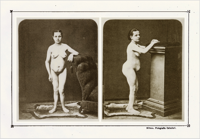
Photographs : 4 mounted albumens on 2 printed leaves, front and side views of the 25 year-old female subject ; anterior
and posterior views of the sectioned uterus.
Photographer : Icilio Calzolari (1833-1906).
Subject : Uterus & adnexa — Caesarean hysterectomy ; Porro's operation.
Notes :
Nella prima tavola le fotografie dell'operata servono a dimostramela configurazione schelčtrica e la nutrizione generale. Nella figura di fronte si vede l'estensione e la direzione della ferita da gastrotomia ela deviazione degli arti inferiori. Nella figura di profilo si riscontra la grave insellatura lombare ed il modo di arcuazione delle coscie.
Nella tavola seconda, la figura superiore rappresenta l'utero amputato, veduto dalla sua superficie anteriore, coll'incisione fatta dal tagliente e la slabbratura accidentale. Si noti l'ubicazione della ferita alla parte inferiore del corpo uterino e sul segmento inferiore, la direzione e forma delle rughe peritoneali. L' ovaja destra si vede pendente al margine destro della matrice, al cui margine sinistro si vede il moncone ovarico e legamentoso.
La figura inferiore mostra l'utero della superficie posteriore: s'osservi il livello del punto di sezione orizzontale posteriore confrontatocolla porzione anterior inferiore di sostanza uterina, che č assai piů basso.
In ambo queste figure si vede il serranodo attorcigliatore del Cintrat colla rispettiva ansa di costrizione , che servě all'amputazione utero-ovarica.—Page 84 (offprint).
These historic photographs represent a 25 year-old primigravid, Madam Julia Cavallini, the first woman to undergo a complete caesarean hysterectomy, succeeding in survival of both infant and mother. Visible in the front and side poses are a marked pituitary dwarfism, rickets, and lordosis (lumbar spondylolisthesis) which distorted the pelvic region of her small frame and made normal delivery impossible. Amputation of the uterus was not Porro's intention, but he was forced into the procedure by an alarming haemorrhage that could not be controlled and the inevitable risk of infection (peritonitis). After removing the child, Porro lifted the uterus from its position in the abdomen and looped the uterine neck opposite the internal os with a wire constrictor (Cintrat's serre-noeud) to stop the bleeding and section the organ. Two additional photographs, taken from above, show the instrument juxtaposed with posterior and anterior views of the specimen uterus. Porro was not the first to attempt the operation—had his patient survived, the procedure would be known today as the Horatio Storer operation (reported by Bixby, 1869, p. 223). It was proposed by others and proven in animal studies conducted at Guy's Hospital by James Blundell in 1823. The skill, the clear-eyed equanimity and depth of scholarship of Porro's report made it an immediate classic and his operation was quickly taken up and improved by surgeons around the world, thereby saving lives and ushering in the era of modern obstetric surgery.