Image source: Google books (uploaded September 2, 2020: »»)
Wiesbaden : Verlag von J. F. Bergmann, 1886.
Journal : Zeitschrift für Ohrenheilkunde und für die Krankheiten der Luftwege ; vol. xvi.
Description : 119–210 p., [1 l.] pl. ; illus: 12 phot. figs., tbls. ; 25 cm.
Photograph : collotype (Lichtdruck). Composite photograph of specimens.
Subject : Labyrinth — Necrosis ; sequestra.
Notes :
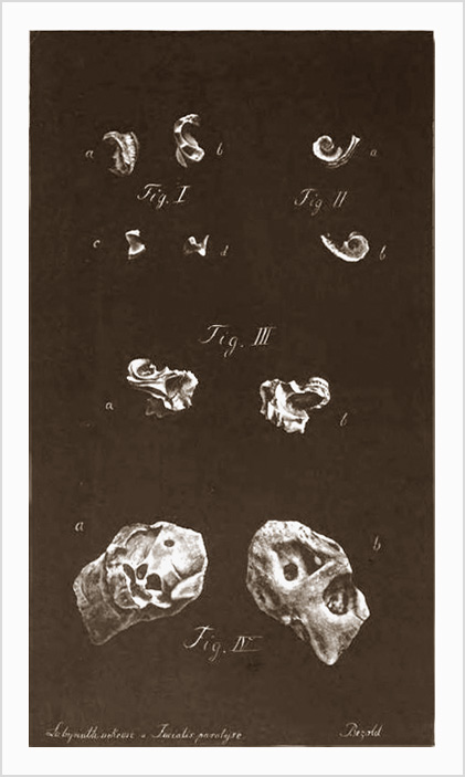
Fig. I. a and b, lower turn of the cochlea, a seen from without, b from within ; c and d, two more small fragments of bone,
the first of which is recognized as belonging to the cochlea. (See history of case No. 2, Rieger).
Fig. II. Lower and middle turns of the cochlea, a seen from without, b from within. (See history No. 3., Blaim).
Fig. III. Lower and middle turn of the cochlea, with a large part of the vestibule and the porus acust. int., a seen from
without, b from within. (See history No. 4., Dallmeyer).
Fig. IV. Sequestrum embracing the whole labyrinth and canalis Fallop., a seen from without, b, from within.
(Case No.5, Stirnweiss).
Adduced from the literature, Bezold tabulated 46 cases of necrotic destruction of the labyrinth with columns of data for age, gender, duration of infection, pain, polypoid growths, vestibular function, hearing tests, sequestra, facial nerve function, extirpation, outcome, and complications. Bezold treated 5 of the 46 patients and provides their histories, illustrated by front and back photos of sequestra removed from 4 of the patients. One of Bezold's histories, fourteen month old Joseph Dallmeyer, is redacted from a clinical report he published earlier in Ärztliches Intelligenz-Blatt (1884, p. 576-577: »»). The analysis revealed nearly twice the number of affected males over females, general onset before age 10, duration up to 20 years, etiology of acute exanthema as a significant indicator, especially in children. Nothing that would surprise otologists, but Bezold's quest is information on necrotic incursion of the Fallopian canal and subsequent paralysis of the facial nerve that might open up new areas of research. In this regard, he records complete data for 35 patients of whom a cohort of 28 suffered extensive necrosis leading to facial paralysis. The data is incomplete for one extraordinary anomaly, Bezold's patient Anna Stirnweiss, who admitted no facial paralysis, even though her sequester encompassed her entire Fallopian canal, exhibited in the photograph (Fig. V). Another patient, Rieger, whose sequester is Fig. I in the photograph, could only raise the corner of her mouth when commanded to raise her eyebrow, both cases leading Bezold into thoughtful commentary on the regeneration of the facial nerve.