Journal : Revue photographique des hôpitaux de Paris ; vol. 4.
Paris : Adrien Delahaye, 1872.
Description : [1 l. pl.], 71-77 p. ; ill.: 1 phot. ; 24.5 cm.
Photograph : 1 mounted albumen.
Subject : Hand — Ulnar nerve; Guyon canal stenosis.
Notes :
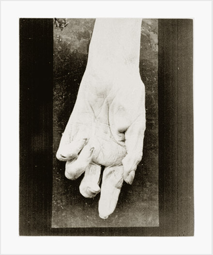
Voici ce que l'examen nécroscopique nous a appris. A l'extrémité inférieure de l'avant-bras gauche, prčs du bord cubital, on observe une cicatrice linéaire, transversale, légčrement sinneuse, représentée sur la Planche IX par une trainée blanche. Elle a son sičge précis entre le pli supérieure et le pli moyen de l'articulation radiocarpienne, commence au-dessus du pisiforme et finit un peu au delŕ de l'axe de l'avant-bras.—Page 71.
From the time of his death in 1921, it has taken nearly a century for a biographical memoir to be written on the life and contributions of Dr. Henri Duret, whose name is forever associated with petechial hemorrhages in the brain stem (O. Walusinski, European Neurology, 2014 »»). The photograph was taken post mortem and represents claw-hand deformity suffered by a 74 year-old woman who died from kidney failure—accidents urémiques. Nerve damage to the subject's wrist occurred fifteen years prior, when she fell onto the head of a broken bottle. I believe this is the first published photograph of ulnar claw.