Journal : Photographic review of medicine & surgery ; vol. 2., no. 4.
Philadelphia : J. B. Lippincott & Co., 1871-72.
Description : pp. 34-36, [1] pl. ; ill.: 1 photo. ; 24 cm.
Photograph : mounted albumen.
Subject : Neck — Encephaloid tumors.
Notes :
NANCY SCHIVELY, aged 55, a native and resident of Chambersburg, Pa., presented herself at the Clinic of the Jefferson Medical College with a tumor of the left parotid region.
The history elicited was as follows :
The tumor first made its appearance twenty-nine years previous, in the form of a small nodule immediately over the left parotid gland (following the extraction of a decayed tooth on the affected side). This increased in size gradually, but slowly, and caused her no material inconvenience until about two years previous to our examination, when it commenced to grow rapidly, and particularly so during the last year, when it attained the enormous size shown in the accompanying photograph.
Although her general health was good, she suffered at times great pain, of a dull, aching character, in the tumor. Her power of mastication and deglutition was also much interfered with.
Upon examination, we found this enormous growth to extend over a space included in a line drawn from the sterno-mastoid process to the median line of the neck antero-posteriorly, and from the horizontal portion of the inferior maxilla to the zygomatic process.
Its measurements were seventeen inches in circumference at the base, and from the parotid gland to the middle of the clavicle twelve and three-quarter inches. The skin was attenuated, with enlargement of the subcutaneous veins. There was no lymphatic involvement in the neighborhood. At several points there were circumscribed elevations of a fluctuating character, which, upon the introduction of the exploring-needle, gave vent to a small quantity of fluid of a chocolate color, which was odorless. At other points it was dense and hard to the touch. It was slightly movable, and manipulation produced pain.
The patient opened the mouth more upon the right than upon the left side. Upon examining the mouth there was found a fluctuating growth, evidently connected with the tumor. There was complete paralysis of the muscles on the left side of the face, with total inability to close the eyelid of that side.
The operation was performed by Professor Pancoast in the following manner :
The patient having assumed the recumbent position, and being placed thoroughly under the influence of ether, an incision was made extending from the lobe of the ear to the inferior margin of the tumor. The superficial and deep fascias were raised, and divided successively on the grooved director. The tumor was firmly held in its bed by bands of fibrous tissue running in various directions, so much so that it was found necessary to raise and divide them separately previous to removal. The parotid gland remained intact, though the anatomy of the neck was thoroughly explored and the muscles and great vessels of the neck exposed to view.
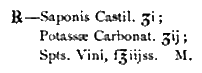
But little hemorrhage followed the operation, it being necessary only to ligate the occipital artery, together with some smaller vessels. The oozing was thoroughly controlled by means of the soap styptic, much esteemed by Professor Pancoast, which is composed of:
The edges of the wound were coapted by means of silk sutures, and the ointment of oxide of zinc was applied as a dressing. The patient had not the slightest untoward symptom, the redundant skin contracting perfectly, and the paralysis of the orbicularis palpebrarum, as well as of the other facial muscles, disappearing gradually.
The patient returned home at the expiration of four weeks, entirely recovered.
The examination of the tumor, made after removal, by my friend Dr. Deal, is as follows :
Upon general examination it was found to be made up of cysts composed of compact masses of grayish matter, bound together by cellular tissue, each cyst containing a small quantity of chocolate-colored fluid. There was evidently no involvement of the parotid gland. The grayish matter of the cysts when subjected to microscopical examination was found to be composed of fibrous matter, the fibres interlaced and forming a dense, compact mass.
Intermingled abundantly with these fibres, and caught in their meshes, were found nuclei and nucleated cells. According to Paget, the less abundant the nuclei the more perfect is the fibrous structure. The basis structure, or stroma, of this specimen was, notwithstanding the abundance of nucleated cells, markedly fibrous. We are taught, however, that "the fibrous tumor sometimes takes on malignant action, its tissues serving as a nidus for the deposition of carcinomatous matter" (Gross); and the appearance of the cells indicated most decidedly that this tumor was encephaloid in its character. This opinion is confirmed by the fact that for twenty-seven years its growth was very slow, while during the last two years it enlarged very rapidly.
There was no appearance of a deposition of either cartilaginous or bony material.