Paris : Librairie Chamerot et Lauwereyns, 1868.
Printed by E. Martinet, Paris, France.
Description : iii, vii, [114] pp. ; ill., [49] plates (part col.), 29 cm
Issued in 12 numbers, 1867-1868.
Photographs : 49 stub bound leaves, each with a mounted albumen.
Photographer : A. de Montméja,
Second Edition (1872 - revised and enlarged) :
Clinique Photographique des Maladies de la Peau, par...
iv, ii, vii, [121] pp. ; ill., [60] plates (part col.), 29 cm.
Third Edition (1882) :
iii-128 pp. ; 60 pl. photo. (woodburytypes).
Subject: Skin Diseases — Atlases.
Cited :
Goldschmidt, Lucien, & Naef, Weston J., Truthful Lens ;
New York, Grolier Club, 1980 :
Both authors had worked at the Hospital Saint Louis in Paris, Hardy being a professor of pathology. The inspiration for the book is acknowledged to have come from an English work seen in 1866, according to Yanul's unpublished research. A second edition was issued under the title "Clinique photographique des maladies de la peau" ; Paris, Lauwereyens, 1872, with 60 photographs, also partly colored.
Notes :
Scholars can browse a first edition of this atlas by accessing the website of Bibliothèque Interuniversitaire de Médecine: BIUM»»
Translating from the introduction, Montmeja writes:
"Dans le courant de l'été 1866, M. Hardy eut connaissance d'essais photographiques faits en Angleterre, et me confia, dès lors, le projet d'étudier avec lui ce nouveau procédé d'iconographie dermatologique. Je commençai par devenir photographe. (…) Les coloris confiés à des mains habiles s'exécutent entièrement sous mes yeux avec la sanction de M. Hardy qui juge en dernier ressort "
"During the course of summer 1866, Mr. Hardy was informed of photographic tests carried out in England, and so he entrusted to me the project to study with him this new process of dermatological iconography. I started by becoming a photographer. The colorings entrusted to skilled hands were executed entirely under my eyes with the sanction of Mr. Hardy who was the judge of final recourse."
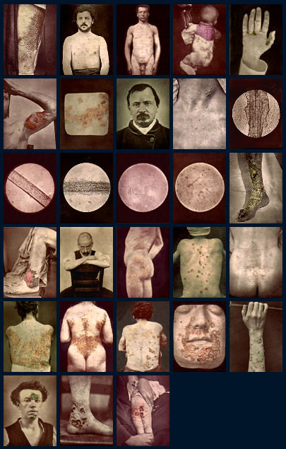
The images vary from book to book and sometimes a completely different model is used. Most of the compositional variations are a slight shift in perspective so it is reasonable to assume that Montmeja was using Disderi's camera fitted with four lenses (and four shutter releases).
The following selection of thumbnails are linked to enlargements:
Because the atlas was issued in parts and consistent with other subscribed atlases of this period, it is not paginated nor are the plates numbered. However, the index does number the plates.
The second edition was enlarged by 12 images which illustrate the following skin disorders: