Journal : Smithsonian Contributions to Knowledge ; vol. xvi .
Washington : Smithsonian Institute, 1870.
Description : 75 p., [16 l.] pl. ; ill.: 36 photos., engrs. ; 33 cm.
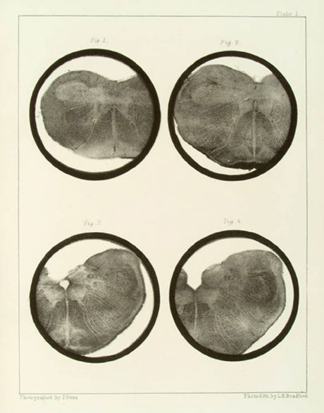
Photographs : 9 leaves of Bradford photolithographs, each with 4 figures — sections of brainstem.
Photographer : John Dean.
Subject : Brainstem — Histology.
Notes :
My friend Dr. Dean, of Boston, U. S., has just sent me some very perfect photographs of sections of the medulla oblongata. These are by far the most perfect photographs of structures from the higher animals that I have seen. ("The Grey Substance of the Medulla Oblongata and Trapezium," by John Dean, M.D., Smithsonian Contributions to Knowledge, 173. -Washington, 1864.) — from page 151, How to Work with the Microscope, by Lionel S. Beale (Philadelphia: Lindsay and Blackiston, 1865).
My friend Dr. Dean, of Boston, U.S., sent me some very perfect photographs of sections of the medulla oblongata, taken with the low powers. These are by far the most perfect photographic illustrations of structures from the higher animals that I have seen. (" The Grey Substance of the Medulla Oblongata and Trapezium," by John Dean, M.D., Smithsonian Contributions to Knowledge, 173- Washington, 1864.) These photographs were also successfully printed by photolithography. Dr. Duchenne, of Boulogne, also obtained some very successful results with anatomical structures, and M. Rouget has employed the same means in the ordinary way and stereoscopically, to illustrate some of his views in minute structure. — from page 233, How to Work with the Microscope, by Lionel S. Beale (London: Harrison, Pall Mall, 1868).
Dr. John Dean's monograph on the histology of the brainstem was selected for publication in volume thirteen of the Smithsonian Contributions to Knowledge (1863), but it was pulled after a fire destroyed the expensive steel engravings that had been prepared for printing illustrations. However, the text blocks were not destroyed and advance copies had been printed. A few copies were bound with albumen photographs and began circulating in 1863. A year later, advance copies illustrated with Bradford photolithographs also began to circulate, issued by the publisher Collins of Philadelphia with the Smithsonian imprimatur. The first edition finally appeared in the 1870 volume of the Smithsonian Contributions to Knowledge, illustrated with replenished engravings of Dr. Dean's drawings and the photolithographs of his negatives. Bradford photolithography was a version of the Poitevin process, improved and patented by Lodowick Harrington Bradford of Boston.
Dr. Dean was called the pioneer of American microscopic studies of the nervous system. His first study on lumbar enlargement of the spinal cord was extracurricular to his medical studies at Harvard and published the year of his graduation, 1860. His major opus on the comparative anatomy of the brainstem left him exhausted, incapacitated by respiratory disease and he was forced to give up his scientific research.